Adenosine Receptor A1
![]()
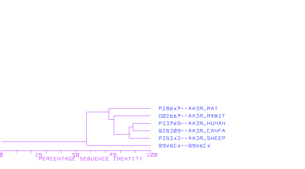
To date, a number of A1 adenosine receptors (A1ARs) has been cloned from a variety of species, including the rat, human, bovine and rabbit (Palmer and Stiles, 1995; Olah and Stiles, 1995). The overall structures of these clones and the expressed protein are remarkably similar in that each encodes a protein of about 326 amino acids, which represents a protein with molecular weight of 36, 000- 37, 000 daltons. Pharmacologically, these receptors are similar to each other, except for bovine A1AR, which has a slightly different agonist potency series. A study by Tucker et al. (1994) suggests that specific amino acids within the transmembrane domain 7, namely Ile- 270 and Thr- 277, are responsible for the species differences in ligand binding to A1 adenosine receptors. This receptor is chiefly linked to the inhibition of adenylyl cyclase activity. However, there is also good evidence for coupling (via G-proteins) to potassium ion channel, an phospholipase C (Palmer and Stiles, 1995; Olah and Stiles, 1995).
Structure- function Relationship
Insight into the structure- function relationships within the A1ARs is being provided by a number of mutation studies and the creation of chimeric receptors.
Mutational Studies
Olah et al (1992) have directly mutated His- 251 in transmembrane domain 6 and His- 274 in transmembrane 7 of the bovine A1 rceptor and replaced these with leucine residues. Subsequent expression and binding studies reveal that mutation of His- 274 resulted in a complete loss of both agonist and antagonist binding.
Work by Townsend- Nicholson had shown that similar mutation in the human A1AR also results in a loss of agonist and antagonist binding, while immunocytochemical analysis with receptor- specific antibodies demonstrated that the receptor was indeed expressed in membrane. This suggests that His- 274 may be very important in binding of agonists and antagonists into the receptor. The replacement of of His- 251 in transmembrane domain 6 results in a fourfold decrease in antagonist affinity, while agonist affinities remain unchanged (Olah et al., 1992).
Townsend- Nicholson and Schofield (1994) have demonstrated that mutation of Thr-277 to alanine in transmembrane domain 7, which lies very close to His- 274, produced a 400-fold decrease in agonist NECA affinity while the affnity of antagonist radioligand (DPCPX) and R- PIA and S-PIA was relatively unchanged
Chimeric Receptors
Additional insights are provided by the creation of chimeric receptors in which various regions of A1ARa and A3ARs are interchanged to create unique receptors that contain components of each of the receptor subtypes.
An interesting chimeric receptor was created in which the sequence of 11 amino acids of the distal portion of the second extracellular loop of the A1AR is placed into A3AR. This replacement results in a chimeric receptor that displays ligand affinities much closer to the A1AR receptors than the A3AR. These data strongly suggest that the second extracellular loop, particularly the distal one third, is important in bindings of ligands into the receptors (Olah et al., 1994c).
Using the chimeric receptor approach, Olah et al., (1994b) have also been able to shown that a 6- amino acid segment of transmembrane domain 5 at the extracellular membrane interface is important in the binding of 5' substituted agonists. For example, the replacement of the transmembrane domain 5 of the A1AR receptor with the analogous region of the A3 receptor results in binding parameters identical to to the wild- type A1 receptor with regard to antagonist binding and the interaction of those agonists that have a substitution on at the N6 position. However, the bonding of 5' substituted agonists is significantly enhanced at the chimeric receptor compared to the wild- type A1 receptor
Thus, multiple regions within the receptor appear to be imortant for both agonist and antagonist bonding, and these regions include transmembrane domains 5, 6 and 7 as well as the second extracellular loop. Further studies are clearly going to define various other region within the transmembrane domain important for agonist and antagonist binding.
| CFGPCR7 | A1-Adenosine Receptor | Canis familiaris | |
| RATA1ADREC | A1-Adenosine Receptor | Rattus norvegicus | |
| RATA1ARA | A1-Adenosine Receptor | Rattus norvegicus | |
| BOVA1ADRE | A1-Adenosine Receptor | Bos taurus | |
| BTARA1 | A1-Adenosine Receptor | Bos taurus | |
| S45235 | A1-Adenosine Receptor | Homo sapiens | |
| HUMADORA1X | A1-Adenosine Receptor | Homo sapiens | |
| S56143 | A1-Adenosine Receptor | Homo sapiens | |
| RABA1AR | A1-Adenosine Receptor | Oryctolagus cuniculus | |
| CPU04279 | A1-Adenosine Receptor | Cavia porcellus | |
| MMU05671 | A1-Adenosine Receptor | Mus musculus | |
| HSA1ADREC | A1-Adenosine Receptor | Homo sapiens | |
| GGU28381 | A1-Adenosine Receptor | Gallus gallus | |
| GGU28380 | A1-Adenosine Receptor | Gallus gallus | |
| AB004662 | A1-Adenosine Receptor | Homo sapiens | |
| AB001089 | A1-Adenosine Receptor | Rattus norvegicus | |
| AF042079 | + | A1-Adenosine Receptor | Rattus norvegicus |
For up to 20 representative sequences

![]()